“Why Does My Knee Keep Hurting When the MRI Is Normal?” ... “Moving Cartilage” Can Now Be Diagnosed [Health Check]
- Input
- 2026-11-18 14:43:45
- Updated
- 2026-11-18 14:43:45
[Financial News] Some patients have pain on the outside of the knee and a catching sensation in the joint, but no clear abnormality shows up on MRI. The cause may be a “hypermobile lateral meniscus,” in which the meniscus moves excessively beyond its normal range. Until now, however, it has been difficult to detect before surgery using MRI.
A research team comprising Professors Kim Jae-gyun and Park Hyeong-jun of the Department of Orthopedic Surgery and Professor Sim Eutteum of the Department of Radiology at Korea University Ansan Hospital has proposed a new assessment measure that may help diagnose hypermobile lateral meniscus (HLM) before surgery. The findings were published in the Korean Knee Society’s international journal, Knee Surgery & Related Research.
The meniscus is tissue in the knee joint that absorbs shock and helps maintain joint stability. When the lateral meniscus on the outer side of the knee moves abnormally, it can cause not only pain and a catching sensation but also locking, in which movement is temporarily restricted.
Unlike a typical meniscal tear, this condition can occur without clear damage to the meniscus itself. Instability when moving the knee is a key feature, but MRI has limitations for diagnosing it accurately because images are taken while the knee is still. If symptoms persist, doctors use arthroscopy to directly observe the abnormal movement of the meniscus and, if needed, suture it to surrounding tissue to restore stability.
Focus on the “fascicles” that hold the meniscus in place; score them from 0 to 2 based on their condition
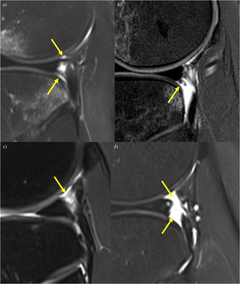
The research team focused on the “popliteomeniscal fascicle (PMF),” which connects the lateral meniscus to the popliteus tendon. The fascicle helps hold the lateral meniscus in place and prevent it from moving excessively when the knee moves.
Until now, assessments have mainly involved measuring the distance between the lateral meniscus and the popliteus tendon or checking for abnormalities in surrounding fascicles, among other factors, in isolation. The team sought a new assessment method to address the limitations of these existing approaches.
The team compared and analyzed preoperative MRI scans from 106 cases of knee arthroscopic surgery performed between December 2013 and December 2024: 53 cases in which hypermobile lateral meniscus was confirmed and 53 in which it was not.
Professor Sim Eutteum explained, “We analyzed various measures, including not only the distance between the lateral meniscus and the popliteus tendon but also the area of the space and tissue characteristics visible on MRI. In particular, we assessed the superior and inferior popliteomeniscal fascicles separately and scored them, aiming to evaluate the condition of the supporting structures around the meniscus more systematically and objectively.”
The research team assessed the fascicles’ continuity and signal brightness on MRI, among other features. They assigned 1 point when a fascicle was clearly connected in a normal configuration, 0.5 points when the connection was incomplete or the imaging showed abnormal features, and 0 points when the fascicle was not visible. They then added the scores for the superior and inferior fascicles to calculate a combined score from 0 to 2.
No difference in distance or area; only the “fascicle score” was associated with the condition—specificity of 86.8%
The analysis found no statistically significant difference between the two groups in the distance between the lateral meniscus and the popliteus tendon or in the area of the space. By contrast, the combined score for fascicle condition was significantly associated with hypermobile lateral meniscus. The less clear the fascicle connections—or the less visible the fascicles—the lower the score and the stronger the association with hypermobile lateral meniscus.
Using a combined score of 1.0 or lower as the cutoff, sensitivity was 58.5%, specificity was 86.8%, and accuracy was 72.6%. In other words, the measure identified about 6 out of 10 patients who had the condition and correctly distinguished about 9 out of 10 who did not.
Professor Park Hyeong-jun said, “Hypermobile lateral meniscus can be difficult to diagnose because the meniscus may in fact be unstable even when no clear abnormality is visible on MRI. The integrated assessment method proposed in this study showed a closer association with the condition than the conventional distance-measurement approach, and we expect it to offer a useful clue to support accurate diagnosis before surgery.”
Professor Kim Jae-gyun said, “This study is significant because it proposes a new approach to comprehensively assessing the condition of the supporting structures around the meniscus on MRI. However, since it is difficult to confirm or rule out the condition using this measure alone, patients’ symptoms and physical examination findings should also be considered. We will continue to validate its clinical usefulness through further research.”
[email protected] Jeong Myeong-jin, medical reporter Reporter