“Went to the hospital for a cough”... ‘Wriggling creature’ found near the lungs of a 38-year-old woman
- Input
- 2026-11-12 19:00:00
- Updated
- 2026-11-12 19:00:01
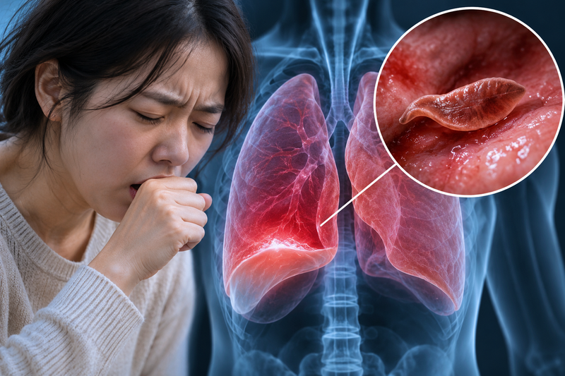
[Financial News] The cough persisted, and her chest felt tight. The chest (pleura) of a 38-year-old woman who thought she had a simple respiratory illness kept filling with fluid for no apparent reason. A blood test showed antibodies to parasites, and she even tried taking antiparasitic medication, but to no avail. In the end, doctors made an incision in her chest and inserted an endoscopic camera. They were stunned by the bizarre sight captured in front of the lens: a living creature clinging tightly to lung tissue and writhing like a wave. It was a lung fluke.
On the 7th (local time), Indian outlet The Times of India reported an unusual case of paragonimiasis in a 38-year-old woman from China, documented in the international journal Infection, Genetics and Evolution.
The problem began with a buildup of fluid in the membrane around the lungs, a condition known as pleural effusion. Her lungs could not expand properly, causing shortness of breath, and she had to be hospitalized twice within six months. During her first hospital stay, blood tests detected antibodies to the Japanese schistosome and sparganum, so she was given an antiparasitic drug (praziquantel), but her symptoms returned. The limitation of blood tests—cross-reactivity between parasites (false positives)—meant doctors could not identify the precise target.
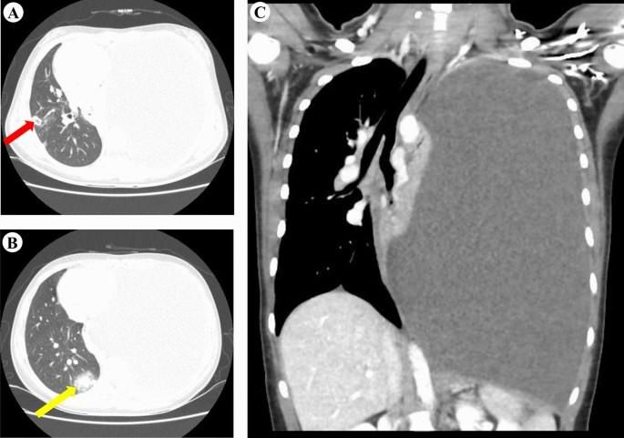
In the end, the medical team decided to perform a thoracoscopy to look directly inside her chest. The camera, aimed near the ribs at the back of her right chest, captured a shocking scene. It was not ordinary inflammation or pus. It was a ‘live parasite,’ contracting and stretching its body as it writhed in waves, much like intestinal peristalsis.
After removing the worm, doctors cross-checked it under a microscope and with metagenomic next-generation sequencing (mNGS), confirming that it was a lung fluke (Paragonimus). According to the researchers, this is the first reported case in the world of a live adult lung fluke being vividly captured inside the chest by a thoracoscopic camera.
Lung flukes are foodborne parasites that usually enter the human body when people eat undercooked freshwater crabs, crayfish or similar foods. The larvae leave the digestive tract, pierce the intestinal wall and diaphragm, and make their way into the thoracic cavity, causing inflammation and fluid to build up. Because the symptoms resemble those of tuberculosis, pneumonia and even lung cancer, the infection is often misdiagnosed.
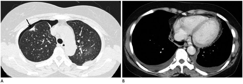
After identifying the exact culprit, doctors again administered praziquantel to target the lung fluke. The pleural effusion that had tormented the woman so severely did not recur over the following four months, and she was declared cured.
The researchers warned, “If unexplained pleural effusion and coughing keep recurring, people should consider whether raw freshwater crustaceans have been eaten and whether there may be a parasitic infection,” adding, “If blood tests do not provide a clear diagnosis, direct endoscopy and genetic analysis may be decisive.” Inside the chest of the 38-year-old woman, who had thought it was just a common cold, a tenacious unwelcome guest was alive and breathing—a presence no one could have imagined before a thoracoscopic camera was inserted.
[email protected] Jeon Sang-il Reporter