“The same red spot, but one is cancer?” Prediction model developed to distinguish Bowen’s disease, which can be hard to tell apart even on biopsy [Health Check]
- Input
- 2026-10-21 08:48:23
- Updated
- 2026-10-21 08:48:23
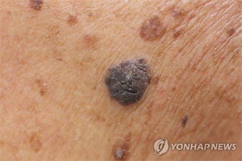
[Financial News] A study has found that whether a lesion develops on the face or scalp, which receive substantial sun exposure, and whether there is UV damage to the surrounding skin may be key clues in distinguishing skin cancer from precancerous lesions.
A research team led by Professor Park Ji-hye of the Department of Dermatology at Samsung Medical Center and Professor Kim Jun-young of the Department of Dermatology at Kyungpook National University Hospital developed a diagnostic prediction model to distinguish Bowen’s disease from bowenoid actinic keratosis by analyzing their clinical and pathological features together.
Bowen’s disease is a skin cancer in which cancer cells are confined to the epidermis, the outermost layer of the skin, and is considered a so-called “carcinoma in situ.” Bowenoid actinic keratosis is a precancerous lesion that may progress to skin cancer. Both conditions can appear as red spots or scaly patches.
The problem is that the two conditions look similar not only in appearance but also under biopsy. There are also not yet sufficiently established consensus criteria for clearly distinguishing them.
Accurate differentiation determines the course of treatment. For Bowen’s disease, a cancer, surgical treatment such as wide excision or Mohs micrographic surgery is considered first. By contrast, bowenoid actinic keratosis, a precancerous lesion, is generally treated with nonsurgical approaches such as cryotherapy or topical medication.
To address this diagnostic challenge, the research team focused on combining biopsy results with clinical features observed on patients’ skin.
The team comprehensively compared lesion location, the condition of the surrounding skin, UV-related changes in skin tissue, and cellular features seen in biopsies among 253 patients diagnosed with Bowen’s disease (152 patients) or bowenoid actinic keratosis (101 patients) at Samsung Medical Center between June 2018 and June 2023.
The analysis found that the clearest difference between the two conditions was evidence of UV exposure. Lesions developed on areas chronically exposed to sunlight, such as the face or scalp, in 90.1% of patients with bowenoid actinic keratosis, compared with just 36.2% of those with Bowen’s disease. UV-related changes in skin tissue were also observed more often in bowenoid actinic keratosis.
Conversely, biopsies found more abnormally shaped cells in Bowen’s disease. The average number of atypical cells observed across a 3-mm width of the epidermis was 13.68 in Bowen’s disease, more than twice the 5.51 seen in bowenoid actinic keratosis.
Based on these findings, the team developed a prediction model combining lesion location, the condition of the surrounding skin, and biopsy results. When the model was subsequently tested on 207 patients at Kyungpook National University Hospital, it achieved 95.17% accuracy, demonstrating strong diagnostic performance.
The significance of this study lies in its presentation of a new diagnostic strategy to help distinguish the two conditions by combining clinical features observable during patient care with pathological information.
Professor Park Ji-hye said, “Bowen’s disease and bowenoid actinic keratosis look similar, but their treatment approaches differ, so accurate differentiation is important. The significance of this study is that it shows how clinical information from skin lesions can be used alongside biopsy information to help distinguish the two conditions.”
The team added, however, that because the model was developed and validated using patients from two medical institutions in Korea, further validation involving patients from diverse ethnicities and regions will be needed. The findings were published in the latest issue of the international journal The Journal of Dermatology.
[email protected] Medical Correspondent Jung Myung-jin Reporter