"My eyes seem a little strange"... Retinal Risks Rise with the Severity of Myopia
- Input
- 2026-09-22 11:24:29
- Updated
- 2026-09-22 11:24:29
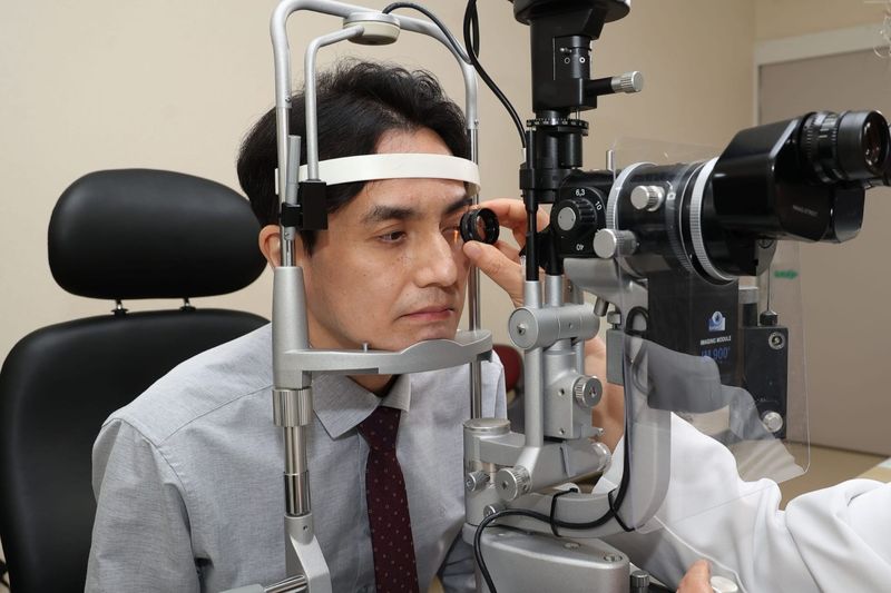
[Financial News] People with myopia, particularly high myopia, should be alert to the risk of retinal detachment. Even when vision is corrected, the eye's elongated structure caused by myopia remains unchanged, so the risk of retinal disease does not disappear.
Kim Ye-ji, a specialist at the Retina Hospital of Kim's Eye Hospital, stressed, "Retinal detachment is not a disease limited to a specific age group and can occur even in relatively young people. In particular, those with high myopia should pay attention to their retinal health. If they experience unusual changes such as a sudden increase in floaters, flashes of light or a curtain-like obstruction in their field of vision, it is important to seek ophthalmic care without delay to protect their eyesight."
The early symptoms of retinal detachment are easy to overlook. If the condition progresses to the macula, which is responsible for central vision, visual recovery may remain limited even after surgery. Regardless of age, it is therefore important not to ignore suspicious symptoms such as sudden changes in the field of vision.
The retina is a thin layer of neural tissue located inside the eye that receives light and transmits visual information. Retinal detachment occurs when the sensory retina separates from the underlying retinal pigment epithelium. There are several types, but the most common, rhegmatogenous retinal detachment, occurs when liquefied vitreous passes beneath the retina through a hole or tear. If the detachment persists, photoreceptor function can be damaged, potentially leading to permanent vision loss or blindness in severe cases.
According to statistics from the Health Insurance Review & Assessment Service (HIRA), the number of patients treated for retinal detachment and retinal tears in 2024 was approximately double the 2014 figure. Patients in their 20s through 50s accounted for 60.1% of the total. In particular, the number of patients in their 50s was similar to that of patients in their 60s, while more than 20,000 patients in their 40s received treatment. This shows that retinal detachment and retinal tears are also being diagnosed relatively frequently among people in their 40s and 50s.
Structural Eye Risks That Persist Despite Vision Correction
Myopia is one of the main risk factors for rhegmatogenous retinal detachment in relatively young people, and the risk is known to be especially high in those with severe myopia. According to the Korean Ophthalmological Society, people with myopia are more likely to develop retinal detachment than those without it, and the risk increases as myopia becomes more severe. As myopia progresses, the eye's axial length—the distance from the front to the back of the eye—increases. This can thin the retina and create conditions in which peripheral retinal degeneration and retinal tears are more likely to occur.
High myopia is therefore not merely a vision-correction issue; it can also be a risk factor for retinal disease. Corrective measures such as glasses, contact lenses, LASIK or LASEK can improve vision, but they do not eliminate structural features of the eye, including the elongated axial length caused by myopia. People should therefore bear in mind that seeing well after vision correction does not mean that the risk of myopia-related retinal disease has disappeared.
In addition to high myopia, various factors can increase the risk of retinal detachment. These include eye trauma, a history of ophthalmic surgery such as cataract surgery, and a history of retinal tears or detachment in the other eye or a family history of the conditions.
The treatment method and visual prognosis for retinal detachment can vary depending on when it is detected. When a retinal tear is present without detachment, laser photocoagulation or cryotherapy may be performed when necessary, taking into account the tear's shape, symptoms and the condition of the surrounding retina. This can reduce the risk of progression to retinal detachment. Once detachment has developed, however, surgical treatment is usually required, with the appropriate procedure determined by the extent and location of the detachment.
Whether the macula is involved is particularly important for visual prognosis. Located at the center of the retina, the macula is responsible for sharp central vision. If retinal detachment progresses to the macula, vision may not fully recover even if surgery reattaches the retina. Detecting the condition and providing appropriate treatment before the macula is affected is therefore crucial for preserving vision.
Watch for Floaters and Flashes That Are Easy to Miss Because They Cause No Pain
Retinal detachment often causes no pain, making it important not to overlook changes in the eyes. People should undergo an eye examination if floaters—such as flying specks, black dots or thread-like shapes—suddenly appear or increase, or if flashes of light newly appear in dark surroundings. Immediate ophthalmic care is necessary if part of the field of vision appears obscured as if by a curtain. People with high myopia are also advised to have regular fundus examinations to monitor the condition of the retina, even when they have no noticeable symptoms.
[email protected] Jung Myung-jin, medical specialist Reporter