A New Device Enables More Accurate Diagnosis of Dense Breasts, Common Among Korean Women
- Input
- 2026-09-14 09:12:25
- Updated
- 2026-09-14 09:12:25
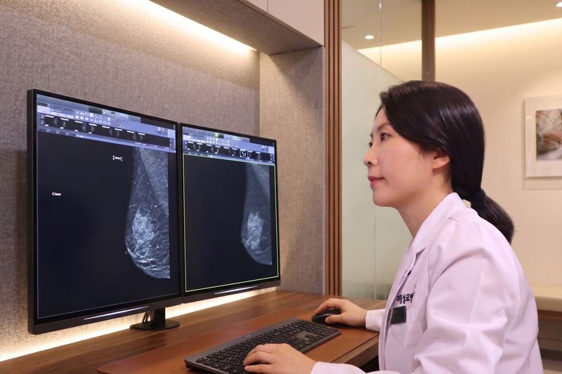
[Financial News] Daerim St. Mary's Hospital has introduced contrast-enhanced mammography (CEM) to diagnose breast diseases more precisely. CEM combines the convenience of conventional mammography with the high sensitivity of MRI. The Ministry of Health and Welfare (MOHW) designated it a new medical technology last February.
CEM combines the convenience of conventional mammography with the high sensitivity of MRI. In February, MOHW designated it as a new medical technology that can be used to diagnose patients with suspicious breast lesions or those requiring follow-up monitoring.
The key feature of CEM is that it adds information on the breast's response to a contrast agent to the structural information visible on conventional mammograms, enabling a more precise evaluation of lesions.
While conventional mammography examines structural changes such as the size and shape of breast lesions and microcalcifications, CEM uses the tendency of contrast agents to accumulate in the blood vessels newly formed by cancerous tissue. This provides additional information about a lesion's blood-flow characteristics and activity.
The examination begins with an intravenous injection of an iodinated contrast agent. The breast is then compressed in the same manner as during conventional mammography. Low- and high-energy X-ray images are acquired consecutively and compared, making lesions that respond to the contrast agent appear more clearly.
These features make CEM useful for diagnosing patients with dense breast tissue. In dense breasts, both normal glandular tissue and lesions appear bright, which can hide lesions within normal tissue. South Korean women have a high prevalence of dense breasts, making diagnosis more challenging in clinical practice.
For patients, CEM offers the advantage of obtaining biological information about tumors that would otherwise require an MRI, while undergoing an examination similar to conventional mammography.
A breast MRI requires patients to remain face down for more than 30 minutes, and it can be difficult for patients with implanted metal devices such as pacemakers. By contrast, CEM is performed while standing, and the entire examination takes about 10 minutes.
Lee So-min, director of the Breast Imaging Center, said, "When diagnosing breast cancer, it is important to appropriately select the necessary imaging tests by considering the patient's breast density and the characteristics of the lesion." She added, "CEM is significant because it can detect small lesions at an early stage that may be hidden by dense breast tissue, while also enabling more precise evaluations during follow-up monitoring."
Daerim St. Mary's Hospital plans to use the introduction of CEM as a foundation for incorporating diverse imaging information into breast cancer diagnosis and further strengthening its specialized care capabilities.
Kim Seong-won, Chairman of the Board of Trustees, stated, "As breast cancer diagnosis and treatment technologies are advancing rapidly, specialized comprehensive breast hospitals must actively adopt new medical technologies that benefit patients." He added, "We will continue to accelerate the adoption of new diagnostic and treatment technologies so that more patients can benefit from advanced medical care."
[email protected] Medical Specialist Reporter Jung Myung-jin Reporter