The Binding Site of a Liver Cancer-Related Protein, Offering Clues for New Drug Design, Identified [Unboxing Lab]
- Input
- 2026-09-02 05:56:00
- Updated
- 2026-09-02 05:56:00
Do you remember the excitement of opening a delivery box? In university laboratories, remarkable discoveries that could change our lives are being made even at this very moment. They are simply wrapped in the thick packaging of academic papers. In “Unboxing Lab,” we will skip the complex formulas and theories and bring you only the key findings you want to know. So, shall we open the box? Today’s featured discovery is this study.
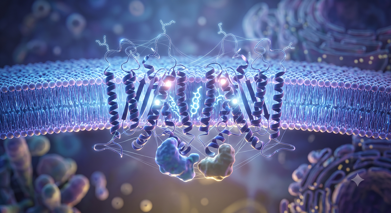
The findings could be used to design treatments for liver diseases such as liver cancer, steatohepatitis, and liver fibrosis. Because the specific site where two TM4SF5 molecules interlock has been revealed, the findings may provide structural clues for identifying therapeutic substances that regulate this binding.
■Tracking Molecular Movements with a Supercomputer
TM4SF5 is a protein that crosses the cell membrane four times. Because cell membrane proteins do not dissolve easily in water, it is difficult to isolate them and determine their structures using conventional experimental methods. To overcome this limitation, the research team used AlphaFold 3, an AI technology for predicting protein structures.
The team generated 100 models of the structure formed by two TM4SF5 proteins binding together and selected the most likely structures. It then used a supercomputer to recreate an environment similar to an actual cell membrane and observed the proteins’ movements for one microsecond, or one-millionth of a second.
■Binding Wavers as the Distance Between Proteins Widens
According to the simulations, the distance between key amino acids at the contact site of two normal TM4SF5 molecules remained close, averaging approximately 4–5 centimeters divided by 100 million. Three to four cholesterol molecules occupied the binding interface and helped stabilize the structure formed by the two proteins.
Glycosylation, the process by which sugar components attach to the outside of a protein, also affected the stability of the binding structure. By contrast, in mutant proteins in which some key amino acids were replaced with different amino acids, the average distance between the two molecules widened to approximately 7–8 centimeters divided by 100 million. The amount of cholesterol attached to the binding interface also decreased.
The research team verified the computer-based findings through experiments using actual cells. It confirmed that the degree of binding between the two TM4SF5 molecules fell sharply when key amino acids were altered or cholesterol levels inside the cells were reduced. This validated the computational finding that key binding sites and cholesterol are involved in dimer formation.
The team also proposed a structural model suggesting that when two TM4SF5 molecules bind, space may form on the inside of the cell where cancer-related signaling proteins such as FAK and c-Src can bind. By presenting the binding structure and mechanism of TM4SF5 in detail, the study could serve as foundational data for future research into treatments targeting the TM4SF5 binding site.
The findings were published in Volume 87 of the international journal Journal of Advanced Research (JAR) in 2026.
[email protected] Man-ki Kim Reporter