"Eye Exams Instead of Brain Scans"...A Single Photo Can Reveal Dementia Risk [Health Check]
- Input
- 2026-08-28 08:57:19
- Updated
- 2026-08-28 08:57:19
[Financial News] Korean researchers have developed an AI system that can assess dementia risk from a single retinal photo. The model also identifies the retinal regions that formed the basis of its judgment, improving its potential for clinical use.
Dementia is difficult to diagnose with a single test, so doctors combine clinical evaluation, cognitive tests and brain MRI scans. However, cost and accessibility limit its use as a broad screening tool. By contrast, the retina is known as an extension of the central nervous system and a tissue that reflects vascular and neural changes in the brain. Fundus images are drawing attention as a biomarker that could help assess dementia risk.
A joint research team led by Professor Park Sang-min of the Department of Family Medicine at Seoul National University Hospital, Dr. Jang Joo-young of Zymed, and researchers Han Chang-ho and Kim Jae-won from the Seoul National University College of Medicine Medical Big Data Research Center and the Department of Biomedical Sciences developed and evaluated an AI model for dementia screening and prediction using 108,008 fundus images from 36,322 health checkup participants at Seoul National University Hospital between 2004 and 2016.
The team defined 1,001 people, or 2,868 images, who were diagnosed with dementia within two years before or after fundus imaging as the dementia group, and built a development dataset by matching them 1-to-4 with a control group without dementia. Using this dataset, the researchers developed 15 AI models by combining five foundation models with three fine-tuning strategies.
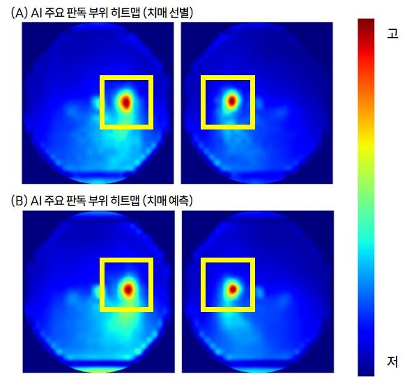
The team then compared the predictive performance of each model, selected the best one, and evaluated its clinical utility and explainability. Explainability was assessed by visualizing, in heatmaps, which parts of the fundus images the AI relied on, and comparing those areas with anatomical retinal structures such as the optic disc and blood vessels.
As a result, the AI model that applied partial fine-tuning to RETFound-MAE, a foundation model pretrained on millions of fundus images, showed the best performance. Its current dementia screening performance reached an AUROC of 0.750, while its prediction performance for future dementia incidence posted a C-index of 0.812, outperforming CAIDE, a conventional clinical dementia risk assessment tool, which recorded an AUROC of 0.624 and a C-index of 0.689.
Even after adjusting for existing dementia risk factors such as age and sex, higher AI scores were associated with a significantly greater risk of both current and future dementia. This suggests that fundus images contain additional information about dementia risk beyond traditional risk factors.
In decision curve analysis, which evaluates the usefulness of prediction models, the AI also scored higher than CAIDE and than both universal screening and no-screening strategies. In addition, when CAIDE was used together with the AI, predictive performance improved, with AUROC rising from 0.749 to 0.774, confirming that the AI model can complement existing dementia risk assessment systems.
Analysis of the AI's decision-making showed high attention to the optic disc and the peripapillary region. Attention to the entire optic disc was 5.72 times higher than that of ordinary surrounding areas, with the highest attention concentrated in the temporal side of the optic disc. High attention was also observed in the papillomacular bundle, which connects the optic disc and the macula, as well as in the peripapillary area. These findings are consistent with retinal regions previously reported in relation to dementia, supporting the possibility that the AI relied on biologically plausible retinal signals.
Professor Park Sang-min of the Department of Family Medicine at Seoul National University Hospital said, "We hope this AI technology, which uses routine screening data without the need for additional high-cost tests, will become a useful support tool for dementia prevention and early intervention strategies."
Dr. Jang Joo-young of Zymed explained, "This study is significant because it evaluated not only the AI's predictive performance but also its explainability and clinical utility, offering an important direction for developing fundus image-based AI biomarkers."
The study was published in the latest issue of the international journal npj Digital Medicine (IF=18.0).
[email protected] Medical Reporter Jung Myung-jin Reporter